Mesenchyme (/ˈmɛsənkaɪm ˈmiːzən-/) is a type of loosely organized animal embryonic connective tissue of undifferentiated cells that give rise to most tissues, such as skin, blood or bone. The interactions between mesenchyme and epithelium help to form nearly every organ in the developing embryo.
Vertebrates
Structure
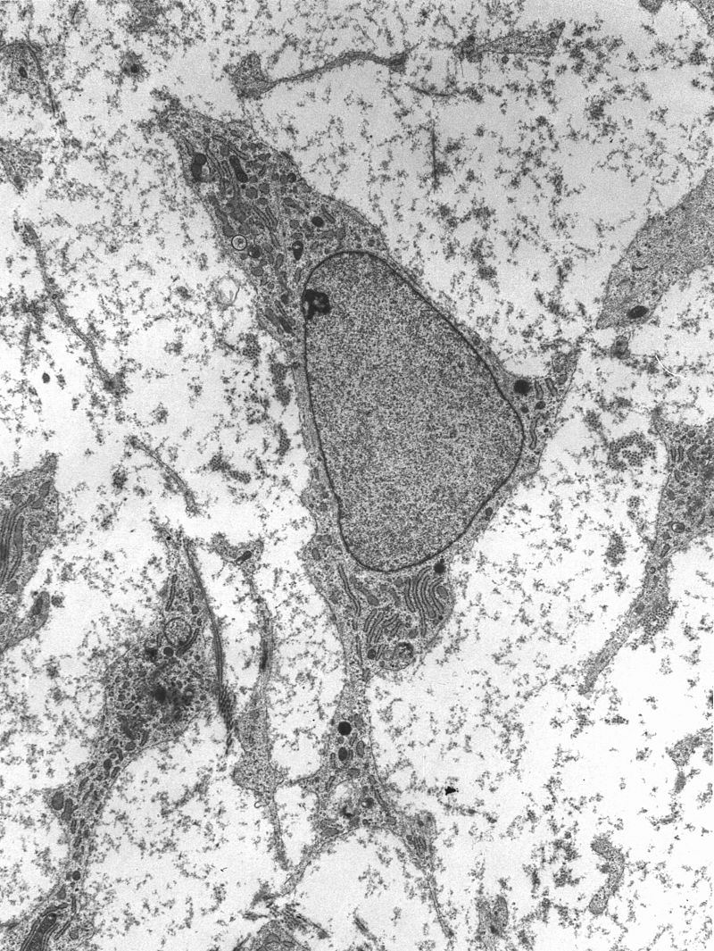
Mesenchyme is characterized morphologically by a prominent ground substance matrix containing a loose aggregate of reticular fibers and unspecialized mesenchymal stem cells. Mesenchymal cells can migrate easily, in contrast to epithelial cells, which lack mobility, are organized into closely adherent sheets, and are polarized in an apical-basal orientation.
Development
The mesenchyme originates from the mesoderm.[6] From the mesoderm, the mesenchyme appears as an embryologically primitive “soup”. This “soup” exists as a combination of the mesenchymal cells plus serous fluid plus the many different tissue proteins. Serous fluid is typically stocked with the many serous elements, such as sodium and chloride. The mesenchyme develops into the tissues of the lymphatic and circulatory systems, as well as the musculoskeletal system. This latter system is characterized as connective tissues throughout the body, such as bone, and cartilage. A malignant cancer of mesenchymal cells is a type of sarcoma.
Epithelial to mesenchymal transition
The first emergence of mesenchyme occurs during gastrulation from the epithelial–mesenchymal transition (EMT) process. This transition occurs through the loss of epithelial cadherin, tight junctions, and adherens junctions on the cell membranes of epithelial cells. The surface molecules undergo endocytosis and the microtubule cytoskeleton loses shape, enabling mesenchyme to migrate along the extracellular matrix (ECM). Epithelial–mesenchymal transition occurs in embryonic cells that require migration through or over tissue, and can be followed with a mesenchymal–epithelial transition to produce secondary epithelial tissues. Embryological mesenchymal cells express Protein S100-A4 (S100A4)[10] also known as fibroblast-specific protein, which is indicative of their shared properties with the migratory adult fibroblasts, and c-Fos, an oncogene associated with the down-regulation of epithelial cadherin. Both formation of the primitive streak and mesenchymal tissue is dependent on the Wnt/β-catenin pathway. Specific markers of mesenchymal tissue include the additional expression of ECM factors such as fibronectin and vitronectin.
Implantation
The first cells of the embryo to undergo EMT and form mesenchyme are the extra-embryonic cells of the trophectoderm. These migrate from the body of the blastocyst into the endometrial layer of the uterus in order to contribute to the formation of the anchored placenta.
Primary mesenchyme
Primary mesenchyme is the first embryonic mesenchymal tissue to emerge, and it is produced from EMT in epiblast cells. In the epiblast, it is induced by the primitive streak through Wnt signaling, and produces endoderm and mesoderm from a transitory tissue called mesendoderm during the process of gastrulation.
The formation of primary mesenchyme depends on the expression of WNT3. Other deficiencies in signaling pathways, such as in Nodal (a TGF-beta protein), will lead to defective mesoderm formation.
The tissue layers formed from the primitive streak invaginate together into the embryo and the induced mesenchymal stem cells will ingress and form the mesoderm. Mesodermal tissue will continue to differentiate and/or migrate throughout the embryo to ultimately form most connective tissue layers of the body.
Source: Wikipedia
Leave a Reply