Giant cell fibroblastoma (GCF) is a rare type of soft-tissue tumor marked by painless nodules in the dermis (the inner layer of the two main layers of tissue that make up the skin) and subcutaneous (beneath the skin) tissue. These tumors may come back after surgery, but they do not spread to other parts of the body. They occur mostly in boys. GCF tumor tissues consist of bland spindle-shaped or stellate-shaped cells interspersed among multinucleated giant cells.
GCF tumors are closely related to dermatofibrosarcoma protuberans (DFSP) and dermatofibrosarcoma protuberans, fibrosarcomatous (DFSP-FS) (also termed fibrosarcomatous dermatofibrosarcoma protuberans) tumors. The World Health Organization (2020) classified these three tumors as different tumors in the category of fibroblastic and myofibroblastic tumors with GCF and DFSP sub-classified as benign but aggressive tumors and DFSP-FS subclassified as a rarely metastasizing tumor. However, The three tumor types may contain areas that have a microscopic histopathological appearance similar to one of the other types. Furthermore, following their surgical resection GCF tumors may recur as DFSP tumors and vice versa and DFSP tumors may recur as DFSP-FS tumors. CGF, DFSP, and DFSP-FS have been regarded as an increasingly aggressive spectrum of related tumors.
Giant cell fibroblastoma tumors are typically treated by surgical resection but have a very high rate of recurrence at the sites of their resection, particularly in cases where all of the tumor has not been removed. Accordingly, wide, complete tumor resections are the recommended treatment for them.
Presentation
As found in one study of 86 individuals, GCF commonly present as single tumors in children less than 10 years old (62% of cases), 10-40 year old individuals (26% of cases), and adults greater that 40 years (12% of cases) (overall median age: 6 years). There is a strong predominance of males in the reported cases of GCF. The tumors are slow-growing, painless, often protuberant, multinodular or polyp-like dermal and subcutaneous tumors masses or plaques (i.e. a lesion that is greater in its diameter than in its depth) that commonly occur on the trunk, upper parts of the arms or legs, or, rarely, the head and neck areas.
Pathology
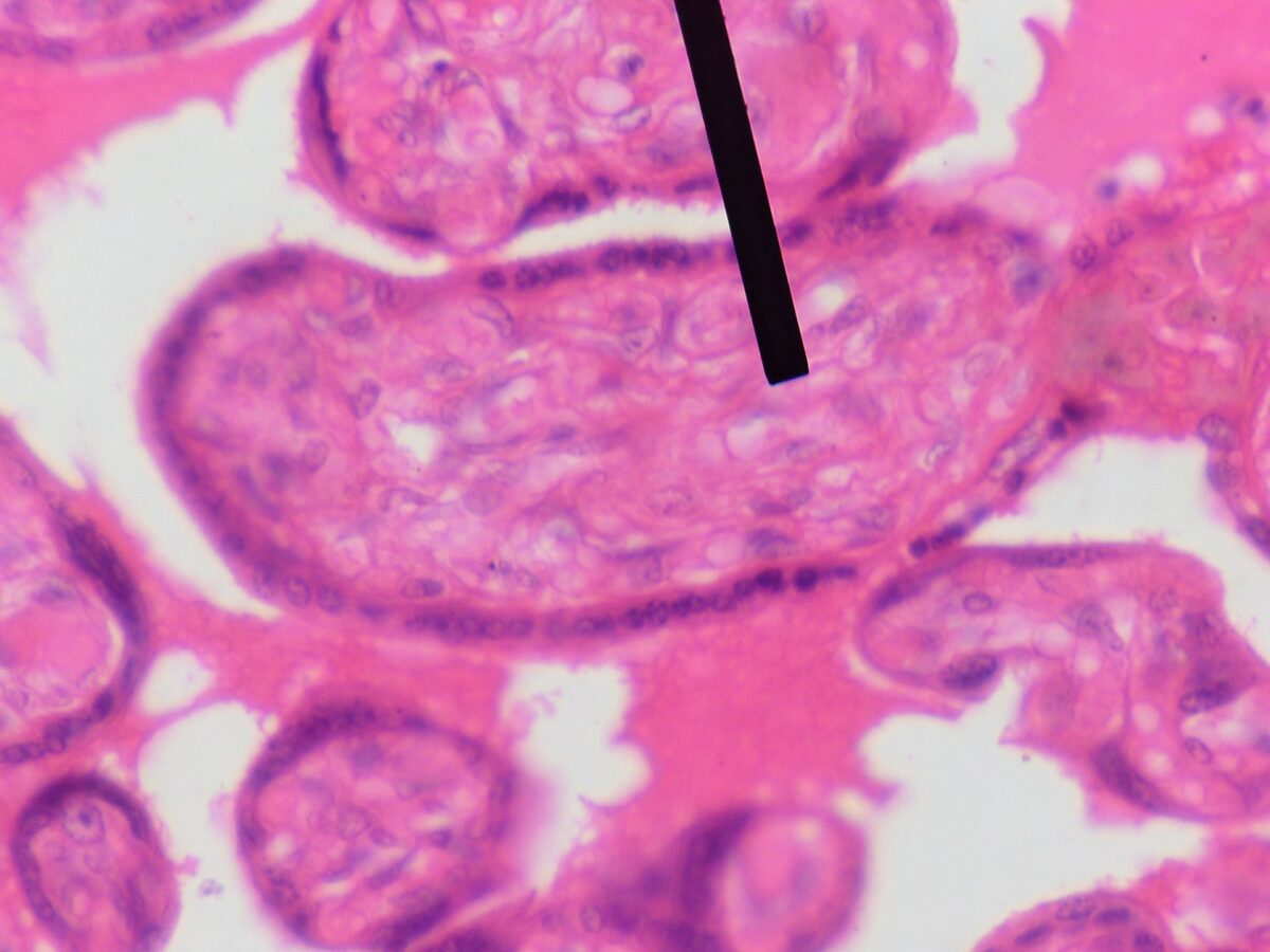
As defined by microscopic histopathology analyses, GCF tumors consist of a few spindle- and/or stellate-shaped cells in a sclerotic (i.e. collagen fiber-rich) background[8] with distinctive dilated blood vessel-like spaces lined by floret-shaped (i.e. small flower-shaped) multinuclear giant cells. The giant cells vary in size and shape with their nuclei often lined-up in wreath-like or lobular formations. The tumors may infiltrate into and through nearby subcutaneous fat tissue, commonly have intralesional hemorrhages and distinctive perivascular onionskin-like lymphocytes, and occasionally contain nodules of smooth muscle-like cells. Some GCF tumors have hybrid characteristics with areas resembling DFSP (e.g. immature-appearing spindle- and/or stellate-shaped cells with abnormally dark nuclei arranged in a monotonous cartwheel or whorled pattern). These hybrid lesions typical have pure GCF-like areas, pure DFSP-like areas, and mixed areas with a gradual or abrupt transition from one to the other. Surgically removed GCF may recur as a DFSP (and vice versa). (DFSP-FS tumors consist of rapidly growing bundles of spindle- and/or stellate-shaped cells with vesicle-containing, abnormally shaped nuclei.
Source: Wikipedia

Leave a Reply