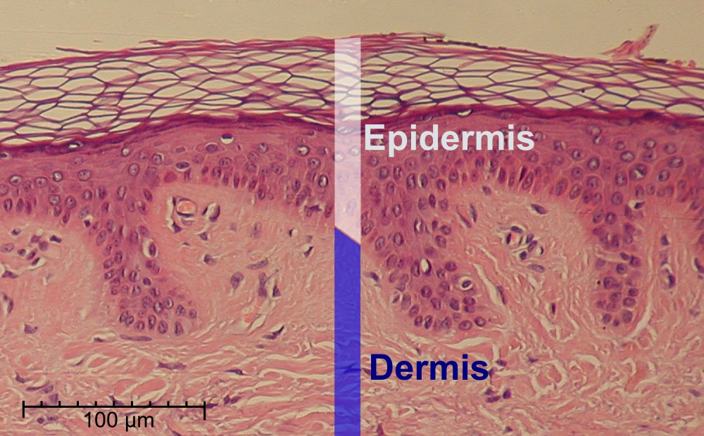
The epidermis is the outermost of the three layers that comprise the skin, the inner layers being the dermis and hypodermis. The epidermis layer provides a barrier to infection from environmental pathogens and regulates the amount of water released from the body into the atmosphere through transepidermal water loss.
The epidermis is composed of multiple layers of flattened cells that overlie a base layer (stratum basale) composed of columnar cells arranged perpendicularly. The layers of cells develop from stem cells in the basal layer. The human epidermis is a familiar example of epithelium, particularly a stratified squamous epithelium.
The word epidermis is derived through Latin from Ancient Greek epidermis, itself from Ancient Greek epi ‘over, upon’ and from Ancient Greek derma ‘skin’. Something related to or part of the epidermis is termed epidermal.
Structure
Cellular components
The epidermis primarily consists of keratinocytes (proliferating basal and differentiated suprabasal), which comprise 90% of its cells, but also contains melanocytes, Langerhans cells, Merkel cells,: 2–3 and inflammatory cells. Epidermal thickenings called Rete ridges (or rete pegs) extend downward between dermal papillae. Blood capillaries are found beneath the epidermis, and are linked to an arteriole and a venule. The epidermis itself has no blood supply and is nourished almost exclusively by diffused oxygen from the surrounding air. Cellular mechanisms for regulating water and sodium levels (ENaCs) are found in all layers of the epidermis.
Cell junctions
Epidermal cells are tightly interconnected to serve as a tight barrier against the exterior environment. The junctions between the epidermal cells are of the adherens junction type, formed by transmembrane proteins called cadherins. Inside the cell, the cadherins are linked to actin filaments. In immunofluorescence microscopy, the actin filament network appears as a thick border surrounding the cells, although the actin filaments are actually located inside the cell and run parallel to the cell membrane. Because of the proximity of the neighboring cells and tightness of the junctions, the actin immunofluorescence appears as a border between cells.
Source: Wikipedia

Leave a Reply